
Rumbo al Centenario de la Microscopía Electrónica de Transmisión
 Descargas: 890
DOI:
https://doi.org/10.56845/terys.v4i1.479Palabras clave:
microscopía electrónica, historia de la microscopía electrónica, microscopía electrónica de transmisión (TEM), Avances en TEMResumen
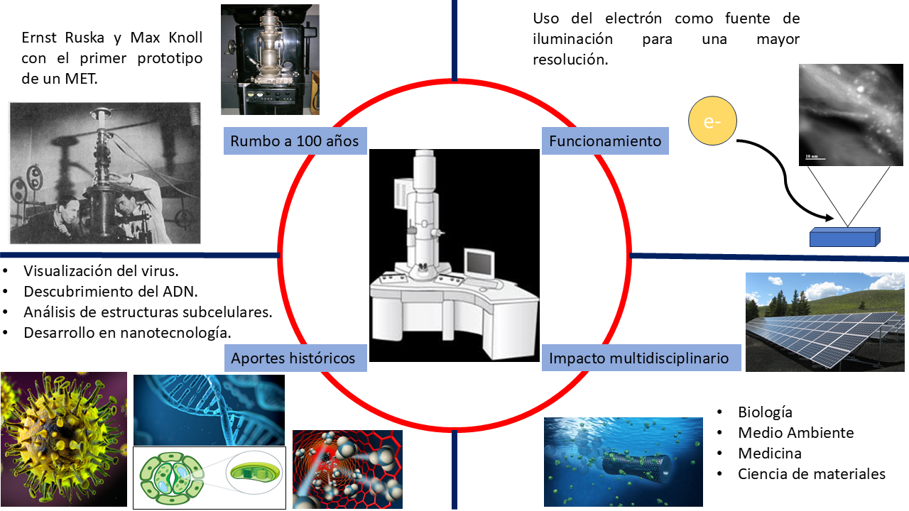
A casi un siglo de su invención, el microscopio electrónico de transmisión (MET) continúa siendo una de las herramientas más poderosas para explorar el mundo invisible. Este artículo presenta un recorrido histórico por los principales hitos que dieron origen a esta tecnología, desde los descubrimientos fundamentales sobre el comportamiento de los electrones hasta la construcción del primer prototipo por Ernst Ruska y Max Knoll en 1931. Se explican también los principios básicos de funcionamiento del MET y se destacan algunos de sus aportes más trascendentes a la ciencia, como la observación de virus, la elucidación de la estructura del ADN, el análisis de ultraestructuras celulares, y los avances en nanotecnología. A través de una narrativa clara y accesible, el texto busca acercar al lector al impacto que ha tenido esta tecnología en disciplinas como la biología, la medicina y la ciencia de materiales, en vísperas de su primer centenario.
Citas
Abbe, E. (1878). Bericht über die wissenschaftlichen Apparate auf der Londoner internationalen Ausstellung im Jahre 1876 (Vol. 1). Vieweg. https://archive.org/details/berichtberdiewi00appagoog
Babarinde, T., & Madyira, D. (2023). Images of Carbon Nanotubes (CNTs) from Transmission Electron Microscopy (TEM) [Micrografías]. Mendeley Data. https://doi.org/10.17632/k8c2bnspxy.1
Bedolla, C. A. (2018). El premio Nobel de Química 2017: microscopía crio-electrónica. Educación Química, 29(1), 3. https://doi.org/10.22201/fq.18708404e.2018.1.63678 DOI: https://doi.org/10.22201/fq.18708404e.2018.1.63678
Bepler, T., Kelley, K., Noble, A. J., & Berger, B. (2020). Topaz-Denoise: general deep denoising models for cryoEM and cryoET. Nature communications, 11(1), 5208. https://doi.org/10.1038/s41467-020-18952-1 DOI: https://doi.org/10.1038/s41467-020-18952-1
Bingham, M., Pesnot, T., & Scott, A. D. (2023). Biophysical screening and characterization in medicinal chemistry. Progress in Medicinal Chemistry, 61–104. https://doi.org/10.1016/bs.pmch.2023.10.002 DOI: https://doi.org/10.1016/bs.pmch.2023.10.002
Busch, H. (1927). Über die Wirkungsweise der Konzentrierungsspule bei der Braunschen Röhre. Arch. Elektrotech. 18, 583–594. https://doi.org/10.1007/BF01656203 DOI: https://doi.org/10.1007/BF01656203
Casciardi, S., Sisto, R., & Diociaiuti, M. (2013). The Analytical Transmission Electron Microscopy: A Powerful Tool for the Investigation of Low‐Dimensional Carbon Nanomaterials. Journal of nanomaterials, 2013(1), 506815. https://doi.org/10.1155/2013/506815 DOI: https://doi.org/10.1155/2013/506815
De Broglie, L. (2021). Research on the theory of quanta (p. L). Montreal: Minkowski Institute Press. https://minkowskiinstitute.org/mip/books/LdeB.html
Deller, M. C., & Rupp, B. (2014). Approaches to automated protein crystal harvesting. Structural Biology and Crystallization Communications, 70(2), 133-155. https://doi.org/10.1107/S2053230X14000387 DOI: https://doi.org/10.1107/S2053230X14000387
Duan, J., Li, J., Chen, G. L., Ge, Y., Liu, J., Xie, K., Peng, X., Zhou, W., Zhong, J., Zhang, Y., Xu, J., Xue, C., Liang, B., Zhu, L., Liu, W., Zhang, C., Tian, XL., Wang, J., Clapham, Zeng, B., Li, Z., & Zhang, J. (2019). Cryo-EM structure of TRPC5 at 2.8-Å resolution reveals unique and conserved DOI: https://doi.org/10.1126/sciadv.aaw7935
structural elements essential for channel function. Science advances, 5(7), eaaw7935. https://www.science.org/doi/10.1126/sciadv.aaw7935
Dubochet, J., Frank, J., & Henderson, R. (2017). The nobel prize in chemistry 2017. Nobel Media AB. https://www.kva.se/app/uploads/2017/10/presskeen17.pdf
Ema, M., Okuda, H., Gamo, M., & Honda, K. (2017). A review of reproductive and developmental toxicity of silver nanoparticles in laboratory animals. Reproductive Toxicology, 67, 149–164. https://doi.org/10.1016/j.reprotox.2017.01.005 DOI: https://doi.org/10.1016/j.reprotox.2017.01.005
Frank, J. (n.d.). The electron microscope: From a sketch in 1931 to reality [Fotografía]. Joachim Frank Lab. Recuperado de https://joachimfranklab.org/the-electron-microscope-from-a-sketch-in-1931-to-reality/
Gatan, Inc. (n.d.). HRTEM images of graphene [Micrografía electrónica]. https://www.gatan.com/resources/media-library/hrtem-images-graphene
Gentile, F., Moretti, M., Limongi, T., Falqui, A., Bertoni, G., Scarpellini, A., Santoriello, S., Maragliano, L., Proietti Zaccaria, R., & di Fabrizio, E. (2012). Direct imaging of DNA fibers: The visage of double helix. Nano Letters, 12(12), 6453–6458. https://doi.org/10.1021/nl3039162 DOI: https://doi.org/10.1021/nl3039162
Gratias, D., & Quiquandon, M. (2019). Discovery of quasicrystals: The early days. Comptes Rendus Physique, 20(7–8), 803–816. https://doi.org/10.1016/j.crhy.2019.05.009 DOI: https://doi.org/10.1016/j.crhy.2019.05.009
Guadalupe, M. U., & Rodríguez-López, J. L. (2007). La nanociencia y la nanotecnología: una revolución en curso. Perfiles latinoamericanos, 14(29), 161-186. https://www.scielo.org.mx/scielo.php?pid=S0188-76532007000100006&script=sci_arttext DOI: https://doi.org/10.18504/pl1429-161-2007
Haguenau, F., Hawkes, P. W., Hutchison, J. L., Satiat–Jeunemaître, B., Simon, G. T., & Williams, D. B. (2003). Key events in the history of electron microscopy. Microscopy and Microanalysis, 9(2), 96-138. https://doi.org/10.1017/S1431927603030113 DOI: https://doi.org/10.1017/S1431927603030113
Iijima, S. (1991). Helical microtubules of graphitic carbon. Nature 354, 56–58. https://doi.org/10.1038/354056a0 DOI: https://doi.org/10.1038/354056a0
Kim, D., Xie, C., Becknell, N., Yu, Y., Karamad, M., Chan, K., Crumlin, E., Norskov. J., & Yang, P. (2017). Electrochemical activation of CO2 through
atomic ordering transformations of AuCu nanoparticles. Journal of the American Chemical Society, 139(24), 8329-8336. https://doi.org/10.1021/jacs.7b03516 DOI: https://doi.org/10.1021/jacs.7b03516
Kim, M., Kim, G. H., Lee, T. K., Choi, I. W., Choi, H. W., Jo, Y., Yoon, Y.J., Kim, J.W., Lee, J. Huh, D., Lee, H., Kwak, S.K., Kim, J.Y., & Kim, D. S. (2019). Methylammonium chloride induces intermediate phase stabilization for efficient perovskite solar cells. Joule, 3(9), 2179-2192. https://doi.org/10.1016/j.joule.2019.06.014 DOI: https://doi.org/10.1016/j.joule.2019.06.014
Klug, A. (1968). Rosalind Franklin and the discovery of the structure of DNA. Nature, 219(5156), 808-810. https://doi.org/10.1038/219808a0 DOI: https://doi.org/10.1038/219808a0
Knoll, M. and Ruska, E. (1932). Das Elektronenmikroskop. Z. Physik, 78, 318–339. https://doi.org/10.1007/BF01342199 DOI: https://doi.org/10.1007/BF01342199
Max Planck Gesellschaft. (n.d.). Images of the Invisible: Max Knoll and Ernst Ruska, 1932 [Fotografía]. Recuperado de https://www.nobel.mpg.de/en/images-of-the-invisible
Novikoff, A. B., Beaufay, H., & de Duve, C. (1956). Electron microscopy of lysosome-rich fractions from rat liver. The Journal of biophysical and biochemical cytology, 2(4), 179. https://www.jstor.org/stable/1603006 DOI: https://doi.org/10.1083/jcb.2.4.179
Novoselov, K. S. (2011). Graphene: materials in the flatland (Nobel Lecture). Angewandte Chemie International Edition, 50(31), 6986-7002. https://doi.org/10.1002/anie.201101502 DOI: https://doi.org/10.1002/anie.201101502
Novoselov, K. S., Geim, A. K., Morozov, S. V., Jiang, D. E., Zhang, Y., Dubonos, S. V., Grigorieva, I. V. & Firsov, A. A. (2004). Electric field effect in atomically thin carbon films. Science, 306(5696), 666-669. https://www.science.org/doi/abs/10.1126/science.1102896 DOI: https://doi.org/10.1126/science.1102896
Palade, G. E. (1955). A small particulate component of the cytoplasm. The Journal of biophysical and biochemical cytology, 1(1), 59.
https://doi.org/10.1083/jcb.1.1.59 DOI: https://doi.org/10.1083/jcb.1.1.59
Porter, K. R., Claude, A., & Fullam, E. F. (1945). A study of tissue culture cells by electron microscopy: methods and preliminary observations. The Journal of experimental medicine, 81(3), 233. https://doi.org/10.1084/jem.81.3.233 DOI: https://doi.org/10.1084/jem.81.3.233
Ruska, E. (1934). Über Fortschritte im Bau und in der Leistung des magnetischen Elektronenmikroskops. Z. Physik, 87, 580–602. https://doi.org/10.1007/BF01333326 DOI: https://doi.org/10.1007/BF01333326
Ruska, E. (1986). The development of the electron microscope and of electron microscopy. Nobel Lecture, The Nobel Foundation. https://doi.org/10.1007/BF01127674 DOI: https://doi.org/10.1007/BF01127674
Shechtman, D., & Blech, I. A. (1985). The microstructure of rapidly solidified Al 6 Mn. Metallurgical Transactions A, 16, 1005-1012. https://doi.org/10.1007/BF02811670 DOI: https://doi.org/10.1007/BF02811670
Stanley, W. M. & Anderson, T. F. (1941). A Study of Purified Viruses with the Electron Microscope. Biological Chemistry, 139, 325-339. https://doi.org/10.1016/S0021-9258(19)51389-8 DOI: https://doi.org/10.1016/S0021-9258(19)51389-8
The Cell Image Library. (n.d.). CIL:7607 – Transmission electron micrograph of rat pancreatic cell showing ribosomes on rough endoplasmic reticulum [Micrografía electrónica]. The Cell Image Library. Recuperado de https://www.cellimagelibrary.org/images/7607
Thomson, J. J. (1897). XL. Cathode rays. The London, Edinburgh, and Dublin Philosophical Magazine and Journal of Science, 44(269), 293-316. https://doi.org/10.1080/14786449708621070 DOI: https://doi.org/10.1080/14786449708621070
Turk, M., & Baumeister, W. (2020). The promise and the challenges of cryo-electron tomography. FEBS letters, 594(20), 3243-3261. https://doi.org/10.1002/1873-3468.13948 DOI: https://doi.org/10.1002/1873-3468.13948
Verhoeven, W., Van Rens, J. F. M., Kieft, E. R., Mutsaers, P. H. A., & Luiten, O. J. (2018). High quality ultrafast transmission electron microscopy
using resonant microwave cavities. Ultramicroscopy, 188, 85-89. https://doi.org/10.1016/j.ultramic.2018.03.012 DOI: https://doi.org/10.1016/j.ultramic.2018.03.012
Von Borries, B., Ruska, E., & Ruska, H. (1938). Bakterien und Virus in Übermikroskopischer Aufnahme: Mit einer Einführung in die Technik des Übermikroskops. Klinische Wochenschrift, 17, 921-925. https://doi.org/10.1007/BF01775798 DOI: https://doi.org/10.1007/BF01775798
Wang, J., Liang, Y., Huang, S., Jin, W., Li, Z., Zhang, Z., Ye, C., Chen, Y., Wei, P., Wang, Y., & Xia, Y. (2022). Conductive graphene coated
carboxymethyl cellulose hybrid fibers with polymeric ionic liquids as intermediate. Carbohydrate Polymers, 280, 119009. https://doi.org/10.1016/j.carbpol.2021.119009 DOI: https://doi.org/10.1016/j.carbpol.2021.119009
Watson, J. D. (2018). ADN. El secreto de la vida (M. Serrano Giménez & I. Cifuentes de Castro, Trad.). Taurus. https://books.google.com.mx/books?hl=es&lr=&id=nshQDwAAQBAJ&oi=fnd&pg=PT23&dq=ADN.+El+secreto+de+la+vida&ots=2J6SQ8rs_P&sig=WfnN1XSg2wI7RDQEIE73yWnZ_qI#v=onepage&q=ADN.%20El%20secreto%20de%20la%20vida&f=false
Williams, D. B., & Carter, C. B. (2009). Transmission Electron Microscopy: A Textbook for Materials Science. Springer. https://doi.org/10.1017/S1431927699990529 DOI: https://doi.org/10.1007/978-0-387-76501-3
Yamane, M., Moriya, S., & Kokuba, H. (2017). Visualization of ceramide channels in lysosomes following endogenous palmitoyl-ceramide accumulation as an initial step in the induction of necrosis. Biochemistry and Biophysics Reports, 11, 174–181. https://doi.org/10.1016/j.bbrep.2017.02.010 DOI: https://doi.org/10.1016/j.bbrep.2017.02.010
Zhang, C., Song, Y., Zhang, H., Lv, B., Qiao, J., Yu, N., Zhang, Y., Di, J., & Li, Q. (2019). Mechanical properties of carbon nanotube fibers at extreme temperatures. Nanoscale, 11(10), 4585–4590. https://doi.org/10.1039/C8NR09637F DOI: https://doi.org/10.1039/C8NR09637F
Descargas
Archivos adicionales
Publicado
Cómo citar
Número
Sección
Licencia
Derechos de autor 2025 César Leyva-Porras, Luis E. Domínguez-Gutiérrez, Raúl García-Torresdey

Esta obra está bajo una licencia internacional Creative Commons Atribución 4.0.
Los autores conservan los derechos de autor de sus trabajos y conceden a la revista Tendencias en Energías Renovables y Sustentabilidad (TERYS) el derecho de primera publicación.
Los artículos se publican bajo la licencia Creative Commons Atribución 4.0 Internacional (CC BY 4.0), que permite compartir y adaptar el material para cualquier propósito, incluso comercial, siempre que se otorgue el crédito adecuado a los autores y a la revista.
Los autores pueden depositar la versión publicada del artículo en repositorios institucionales o páginas personales, siempre citando la publicación original en TERYS.
